Radiological Education in Sengerema

In October, Marieke Vermaat and Mariëlle Mourits, radiologists from Canisius Wilhelmina Hospital in Nijmegen, visited Sengerema Hospital and share their impressions with us:
The adventure began with registration for the Africa Classic Tanzania, a 400 km mountain bike tour around Mt. Kilimanjaro over 6 days, raising funds for Amref Flying Doctors. Shortly thereafter, Bart, a surgeon at CWZ and chairman of SimbaHealth, asked if we could provide radiological education at Sengerema Hospital. We decided to combine both activities and continue our journey after the bike tour.
In Sengerema, we were warmly welcomed by Malouk, one of the tropical doctors, and Erik and Jiska, the founders of Simba Health. We stayed in the container house, a home built from 2 shipping containers on the hospital compound. It exuded a homely atmosphere, making us feel at home. Anastasia and Vivian took excellent care of us, cooking and cleaning so we could focus on teaching.
Radiological examinations available in Sengerema include conventional X-rays and ultrasound. Conventional X-rays involve directing X-rays through the patient to create an image. It forms a two-dimensional picture, especially effective for assessing bones. Structures overlapping each other are less distinguishable, so these photos are taken in two directions for more information. In addition to X-rays, ultrasound examinations are also possible.

Advanced imaging methods like CT scans and MRI, commonly used in CWZ, are unavailable in Sengerema. Adjusting to this difference requires a shift in thinking and practice.
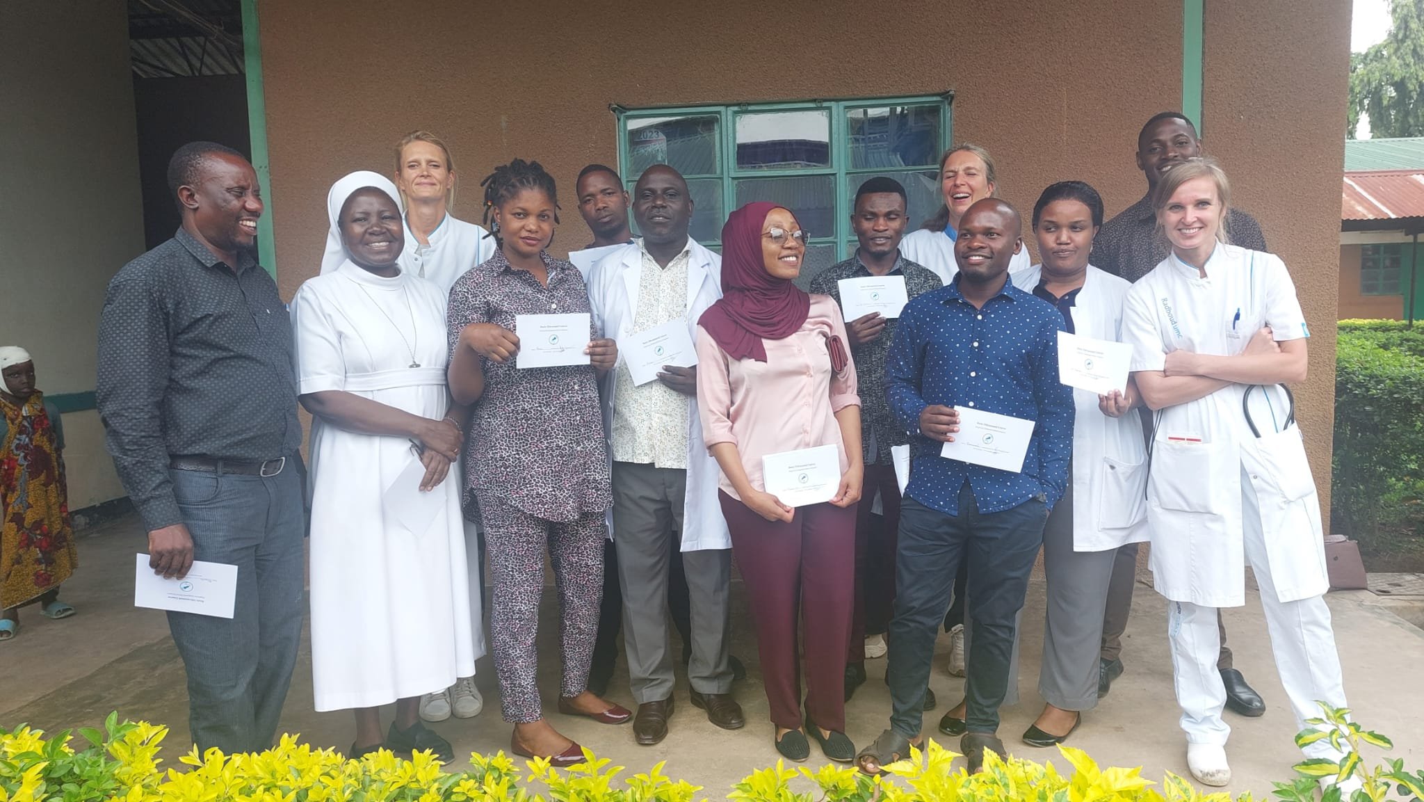
Our daily routine began with morning prayer followed by the handover. We then engaged in educational activities. Initially, we taught the structured assessment of chest and abdominal X-rays, emphasizing a systematic approach to minimize the risk of oversight. After theory lessons, we practiced together with photos taken in the hospital over the past days. Local staff, including doctors and clinical officers, enthusiastically participated, gaining practical insights.

Subsequently, we conducted two basic ultrasound courses for medical doctors and clinical officers. The initial theory covered how ultrasound forms images and how to adjust settings, akin to photography. Practical lessons followed, involving "normal anatomy" ultrasound using the butterfly, an ultrasound head connected to an iPad displaying images.
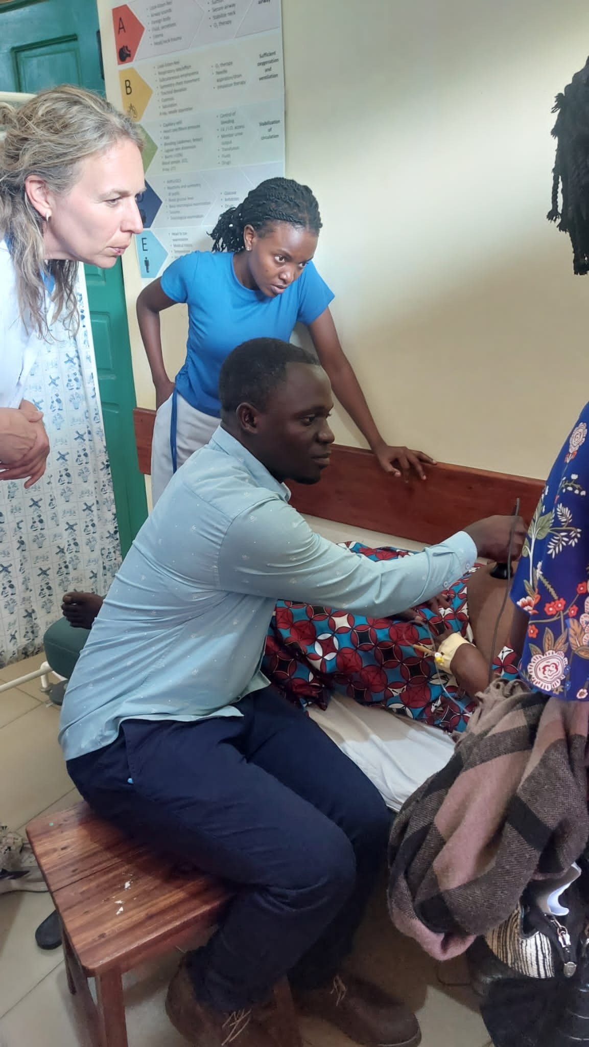
A session on acute abdominal pain and differentiating surgical (e.g., appendicitis) from non-surgical causes (e.g., enlarged liver due to malaria) concluded with practical application, practicing ultrasound on patients in the hospital.

Apart from group lessons, we closely guided the three radiographers during ultrasound examinations and X-ray imaging. Emphasis on structured procedures ensured no organ was overlooked. Additionally, we taught the assessment of deep vein thrombosis using ultrasound, an examination not previously applied in Sengerema.
For X-ray imaging, attention was given to the patient's positioning for optimal image formation.
We also conveyed the importance of objectivity in reporting findings, describing visible abnormalities without drawing conclusions. Explaining situations where patients experience symptoms but no abnormalities appear on the ultrasound can be challenging, especially in Tanzania where trust in ultrasound as an all-seeing eye is significant.
This week exposed us to significant differences between Dutch and Tanzanian healthcare. Socioeconomic disparities impact equal opportunities, and the reluctance to visit hospitals for minor issues in Tanzania means that when patients do come, there's often a serious concern. Complaints persist for a long time, leading to diseases reaching more advanced stages than usual in the Netherlands.
Hygiene and knowledge gaps contribute to healthcare challenges. Homes lack running water, and daily water is fetched from the nearest well or sometimes from Lake Victoria, which may contain parasites. Overcrowded living conditions contribute to the rapid spread of infectious diseases.
The hospital faced shortages of materials and resources, such as echo probes, tubes for draining infections, or needles for IV insertion. The ICU lacked monitors and ventilators, and there were insufficient personnel at the bedside. Much work remains to be done.
We had an unforgettable and intensive week, hoping our contribution enhanced radiological knowledge in Sengerema. Many thanks to Bart, Erik, Jiska, and all hospital staff who made this possible.